In a few years, when researchers investigate how liver cells or heart cells behave, they may very well be using a microscope developed in Denmark.
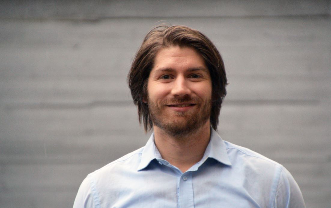
The Novo Nordisk Foundation has awarded Emil Boye Kromann, Associate Professor and Group Leader at the Technical University of Denmark, a grant of more than DKK 13 million over the next 7 years to develop a microscope for studying living tissue in much greater detail than is currently possible.
The development of the new microscope will enable researchers to study cells close to their natural environment and in interaction with other cells. This will greatly advance researchers’ knowledge of how cells behave in the body, how they respond to drugs and what goes wrong when cells develop into cancer cells.
“Receiving such a large grant is amazing. This will provide both continuity and momentum to develop a microscope that many of my colleagues really need,” says Emil Boye Kromann.
Developing a microscope to study miniature organs
Currently, when researchers study cells, such as liver cells, they usually have to study them in a cross-section of dead tissue or isolated cells in a petri dish under a microscope.
These types of microscope studies have been the basis of many medical advances over the past 50 years.
Current microscope technology has some limitations, however, including that cells in a three-dimensional structure surrounded by other cells, as they are in their natural environment, cannot be studied optimally. The deeper in the organ researchers look, the more the images are blurred.
Researchers are therefore seeking new ways to study living cells.
“Today, many of my colleagues use organ-on-a-chip systems to grow a microorgan on a small polymer chip. This microorgan may be derived from intestinal or liver cells, which makes studying how the cells behave while in an organ easier. But my colleagues are still limited by the lack of microscopy techniques to properly study the samples,” says Emil Boye Kromann.
Emil Boye Kromann explains that organ-on-a-chip devices are a very exciting technology that can eventually become an alternative to animal testing. In addition, Denmark is at the forefront of this technology, which also has the perspective of benefiting individualized treatment programmes, in which researchers can grow people’s own cells and study how, for example, their liver will respond to a given drug.
Close collaboration with biologists
Emil Boye Kromann aims to use the NERD project to develop a microscope that can take high-resolution images of organ-on-a-chip systems.
In microscopy, Emil Boye Kromann wants to determine such things as how to optimize the sample’s light signal using the microscope to capture high-resolution images of the events several cell layers deep in the samples.
“The light signal is created as a kind of echo when laser light is beamed into the sample. The idea is to transmit the laser light in a new way so that we can determine more precisely where the echo originates. This will enable us to capture sharper images. In addition, we will try to collect the echo from a larger area of the sample, which enables us to map rapid biological processes, such as the movement of the small vesicles travelling around inside the cells or between them,” explains Emil Boye Kromann.
Bombarding samples with ultraviolet and infrared light
Emil Boye Kromann will also investigate the possibility of studying biological samples with ultraviolet and infrared light.
Today, researchers in biology usually use visible or near-visible light, but information that could be derived outside this spectrum can be hidden, and this may be relevant in further developing microscopy.
“Infrared light is interesting because it does not damage the cells in the same way as visible or near-visible light. Further, it may penetrate deeper into the samples. Ultraviolet light has a shorter wavelength, so it can provide much better image resolution. We will experimentally explore the possibilities of using these near-visible wavelengths in our microscope, but we will also engage in dialogue with researchers working on electromagnetic radiation that is even further away from the visible spectrum,” says Emil Boye Kromann.
The research has only just begun, and it will take up to 7 years for the final microscope to be finished.
During this time, Emil Boye Kromann will work closely with other departments at the Technical University of Denmark (DTU) and will also use part of the grant to include and involve both MSc and BSc students in the project.
Emil Boye Kromann leads a practical course on prototyping at DTU Skylab, where students have access to many workshops and laboratory facilities. He plans to use the course to work with the students to prototype parts of the microscope and possibly seek new perspectives on the project.
“I am really looking forward to this. Overall, the grant means that we can accelerate progress more rapidly than if we had to seek funds for the project and development bit by bit. We can plan far ahead and can link teaching and research so everyone wins,” says Emil Boye Kromann.
The grant awarded to Emil Boye Kromann is part of the Foundation’s new NERD research programme in which the Foundation supports wild and creative research ideas within the natural and technical sciences. The new programme targets ambitious, dedicated researchers who give priority to pursuing their own creative ideas in the laboratory and to maintaining their focus on one theme over the long term to generate new original knowledge. The Foundation has allocated nearly DKK 300 million to NERD.
Further information
Christian Mostrup, Senior Programme Lead, +45 3067 4805, [email protected]


